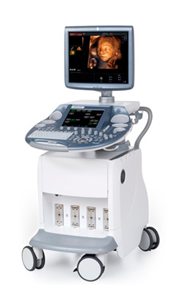
- Access Free Voluson E8 User Manual Voluson E6, E8, and E10 Comparison (Which One to Get. GE Voluson S6, S8, and S10 Ultrasound Transducer Guide. The GE Voluson S6, S8, and S10 ultrasound systems are the result of relentless innovation and improvement that make up the culture.
- GE Voluson E6, E8 Configuration. Last Modified on 2017-05-04 19:01. Fol low the process below to configure your ultrasound system to send data to Tricefy.
The Voluson E6 supports a wide range of 2D and 3D probes to help meet your unique and varied clinical needs. When combined with the advanced system architecture of the Voluson E6, Expert Series probes produce excellent images with ease. Fetal chest and abdomen RIC5-9. Coronal uterus with OmniView RAB6. HD-Flow fetal abdomen RAB6. The Voluson™ E6 is a versatile ultrasound system offering the superior imaging you need today with the flexibility for when your needs evolve. Your entry into the Voluson Expert Series, the Voluson E6, is the perfect choice for a growing practice that demands excellence in patient care.

Product Description

GE VOLUSON E6
Timely access to quality ultrasound is critical in women's healthcare. You need an ultrasound system that gives you flexibility, ease of use, and the speed and reliability to manage a comprehensive range of routine obstetric and gynecological exams. The powerful Voluson E6 system is your answer. It offers an excellent mix of capabilities for effective and efficient care, including extraordinary 2D image quality, outstanding 3D/4D imaging, automation advances, intuitive workflow tools, and a smart ergonomic design.

Product Description
GE VOLUSON E6
Timely access to quality ultrasound is critical in women's healthcare. You need an ultrasound system that gives you flexibility, ease of use, and the speed and reliability to manage a comprehensive range of routine obstetric and gynecological exams. The powerful Voluson E6 system is your answer. It offers an excellent mix of capabilities for effective and efficient care, including extraordinary 2D image quality, outstanding 3D/4D imaging, automation advances, intuitive workflow tools, and a smart ergonomic design.
Voluson E6 is well suited to meet your imaging needs across a range of OB/GYN applications, including assisted reproductive medicine.
Built on the powerful E-Series platform, the Voluson E6 combines advanced probe technology plus innovative hardware and software architecture. Every component works together – processing multiple data points simultaneously in real-time to deliver exceptional images. The Voluson E6 helps you to see more, at earlier stages.
The premium system is designed for OB/GYN, Abdominal, Vascular, Breast, Small Parts, Urology, Cardiology and Orthopedic Applications
Operating Modes
• B-Mode
• M-Mode
• Color M-Mode
• HD Color Flow Mode
• Power Doppler Imaging
• Pulse Wave Doppler (PWD)
• 3D/4D Volume Modes
• Tissue Doppler Imaging
Voluson E10
Features
Ge Voluson E6 User Manual
• High resolution 19' high resolution LCD monitor with DVI Interface
• Touch Screen – 10.4' High Resolution Color LCD screen
• Integrated 500 GB Hard Drive
• 3000 frames standard Cine Memory
• Standard DVD R/W storage
• AO – Automatic Optimization. One-touch image optimization improves contrast resolution, color sensitivity or spectral Doppler depending on the active mode.
• ATO Auto Tissue Optimization
• Coded Harmonic Imaging
• HDlive – This second-generation rendering tool provides exceptional anatomical realism and helps increase depth perception. This imaging capability can help you achieve a deeper understanding of relational anatomy, enrich patient communication and help enhance diagnostic confidence.
• CrossXBeam (Compound Resolution Imaging) helps enhance tissue and border differentiation with a real-time, spatial compounding acquisition and processing technique.
• Speckle Reduction Imaging (SRI) suppresses speckle artifact while maintaining true tissue architecture
• Volume SRI (Speckle Reduction Imaging) (V-SRI) – This enhancement provides a new level of speckle reduction utilizing volume/voxels versus traditional single slice imaging. It helps improve 3D/4D quality in multi-planar studies and rendered mode, and also provides an enhanced smoothing effect on rendered images which helps improve diagnostic confidence.
• HD-Flow uses a bi-directional Doppler feature to help achieve a more sensitive vascular study and reduce overwriting.
• Advanced Volume Contrast Imaging (VCI) with OmniView helps improve your contrast resolution and visualization of the rendered anatomy with clarity in any image plane, even when viewing irregularly shaped structures
• XTD View. Extended field of view allows you to image large organs, typically unable to be seen within a single image.
• Spatio-Temporal Image Correlation (STIC) captures a full fetal heart cycle beating in real time, and the volume can be saved for offline analysis
• Tomographic Ultrasound Imaging (TUI) helps make analysis and documentation of dynamic studies easier with a simultaneous view of multiple parallel slices of a volume data set
• Virtual Convex, enables extended field of view with linear transducers
• SonoNT (Sonography-based Nuchal Translucency) and SonoIT (Sonography-based Intracranial Translucency) – Voluson technologies that help provide semi-automatic, standardized measurements of the nuchal and intracranial translucency as early as 11 weeks. SonoNT helps reduce the inter-and-intra-observer variability that comes with manual measurements, and helps provide you with the reproducibility you demand.
• SonoBiometry – Performs a semi-automatic measurement of the head (both head circumference and bi-parietal diameter), abdomen and femur. This tool can help enhance clinical workflow through helping reduce keystrokes to perform biometry measurements.
• Sonography-based Automated Volume Count follicle (SonoAVC* follicle) is an innovative software program designed to automatically calculate the number and volume of hypoechoic structures from a 3D ovarian volume.
• Sonography-based Volume Computer Aided Display heart (SonoVCAD* heart) is an innovative tool that assists in generating standard views, including the Aortic Arch algorithm, of a fetal heart from a four-chamber view.
• Beta View
• Real-Time automatic Doppler Calcs
• VOCAL – Volumetric Calcs
• Vascular Calcs
• OB/GYN Calcs and tables
• Urological/Renal Calcs
• DICOM 3.0
Transducers
Lagu supernova sayang stafa band lagu. Download Lagu Supernova Sayang Stafa Band Posted on 5/28/2018 by admin Di hey sobatku kita jumpa lagi tempat tongkrongan kita lagi yaitu yang slalu memberi berita terupdate dan info-info terbaru tentang musisi band papan atas yang sedang naik daun maupun musik yang lawas yang masih exis di permusikan Indonesia. Supernova Band Full Album lagu Pop Thn 2000an. Supernova Sayang Lyric video. Download Lagu Terbaru, Gudang Lagu Mp3 Stafa Gratis STAFABAND - Tempat Download Lagu Terbaru, Gudang Lagu Mp3 Gratis 2018, Download Mudah, Cepat dan Gratis. Download Lagu Supernova Sayang Stafa Band 3/15/2018 / Comments off LIRIK LAGU Sayang Kamu telah mengisi lubuk hatiku Jauh dalam relungku Pernahkah kau merasakannya Cinta kini hanya engkau yang bisa Buat ku merasa bahagia Apa kau pun merasakannya Dan memeluknya Ku menangis tertahan Ingat kau tak lagi bersamaku Ku teriris terdiam Berharap. Download Lagu Supernova Sayang Stafa Band On 5/5/2018 By admin In Home LIRIK LAGU Sayang Kamu telah mengisi lubuk hatiku Jauh dalam relungku Pernahkah kau merasakannya Cinta kini hanya engkau yang bisa Buat ku merasa bahagia Apa kau pun merasakannya Dan memeluknya Ku menangis tertahan Ingat kau tak lagi bersamaku Ku teriris terdiam Berharap.
4C-D Convex, M6C-D Matrix Convex, AC1-5-D Convex, SP10-16-D Linear, 9L-D Linear, 11L-D Linear, IC5-9-D Microconvex, PA6-8-D Sector, RAB2-5-D Volumetric Convex, RAB4-8-D Volumetric Convex, RAB6D Volumetric Convex, RIC5-9-D Volumetric Endocavitary, RNA5-9-D Volumetric Microconvex, RSP6-16-D Volumetric Linear
GE's Voluson line is arguably the most popular women's health ultrasound system line on the market. The current GE Voluson Expert series is no different. With the line of BT16 and newer Voluson E6, Voluson E8, and Voluson E10 you can expect world-class imaging at high speeds from each model. You can even 3D print from the newest Voulson systems! And by using the Voluson E6, Voluson E8, and Voluson E10 transducer guide, you can better understand these systems to more efficiently shop for the GE probes they require.
Use the Voluson E6, Voluson E8, and Voluson E10 transducer guide below for reference and when you find the ultrasound probe you need, contact us by filling out a form or call 317.204.3012.
At Probo Medical, we buy, sell, and repair ultrasound probes and stock systems from industry-leading OEM's. Our mission is to deliver the perfect probes and systems at a wholesale cost to every customer, every time.
Ditch witch 1030 parts manual. Summary of Contents for Ditch Witch 1030 Page 1: Service 1030/1230 - SERVICE SERIAL NUMBER RECORD SERVICE SERIAL NUMBER RECORD Record serial numbers and date of purchase in spaces provided. Serial number plate is mounted to frame behind right wheel.
View our full list of ultrasound transducer guides for more options. You can also visit our partner company Providian to read a GE ultrasound buyers guide or visit our partner Ultrasound Supply to shop GE ultrasounds, including the Voluson E Series.
Voluson E6, E8, E10 Transducer Guide
| Probe Type | Probe Name | Description | FOV | Application | Bandwidth |
|---|---|---|---|---|---|
| Small Parts - 2D | |||||
| SP10-16-D | Small Parts, Breast Peripheral Vascular, Pediatrics, Musculoskeletal | 33.7mm | Small Parts, Breast Peripheral Vascular, Pediatrics, Musculoskeletal | 7-18 MHz | |
| 11L-D | Small Parts, Breast Peripheral Vascular, Pediatrics, Musculoskeletal | 37.4mm | Small Parts, Breast Peripheral Vascular, Pediatrics, Musculoskeletal | 4-10 MHz | |
| 9L-D | Small Parts, Peripheral Vascular, Pediatrics, Musculoskeletal | 43.0mm | Small Parts, Breast Peripheral Vascular, Pediatrics, Musculoskeletal | 3-8 MHz | |
| ML6-15-D | Small Parts, Breast Peripheral Vascular, Pediatrics, Musculoskeletal | 49.6 mm | Small Parts, Breast Peripheral Vascular, Pediatrics, Musculoskeletal | 4-13 MHz | |
| Abdominal - 2D | |||||
| 4C-D | Wide Band Convex Transducer | 58° Wide 81° (only E8 Expert) | Abdomen, Obstetrics, Gynecology | 2-5 MHz | |
| C1-5-D | Wide Band Convex Transducer | 69° Wide 113° (only E8 Expert) | Abdomen, Obstetrics, Gynecology | 2-5 MHz | |
| C4-8-D | Wide Band Convex Transducer | 75° Wide 95° (only E8 Expert) | Abdomen, Obstetrics, Gynecology, Pediatrics, Urology | 2-8 MHz | |
| AB2-7-D- | Wide Band Convex Transducer | 80° Wide 107° (only E8 Expert) | Abdomen, Obstetrics, Gynecology, Pediatrics, Urology | 2-8 MHz | |
| M6C | Wide Band Convex Transducer with Active Matrix Array Technology | 60° Wide 84° (only E8 Expert) | Abdomen, Obstetrics, Gynecology, Pediatrics, Urology | 1-7 MHz | |
| Phased Array-2D | |||||
| S4-10-D | Wide Band Phased Array Transducer | 95° | Small Parts, Cardiology, Pediatrics | 4-9 MHz | |
| PA 6-8-D | Wide Band Phased Array Transducer | 90° | Small Parts, Cardiology, Pediatrics | 4-10 MHz | |
| 3Sp-D | Wide Band Phased Array Transducer | 90° | Cardiology, Obstetrics, Abdomen, Neurology, Pediatrics | 1-5 MHz | |
| Endocavity - 2D | |||||
| IC 5-9-D | Wide Band Endocavitary, Micro-convex Array Transducer | 146° Wide 179° | Obstetrics, Gynecology, Urology | 4-9 MHz | |
| MicroConvex - Real Time 4D | |||||
| RNA5-9-D | Wide Band Convex Volume Transducer | 116°, V116° x 90° Wide 114°, V 114° x 90° (only E8 Expert) | Abdomen, Small Parts, Cardiology, Obstetrics, Pediatrics | 3-9 MHz | |
| Abdominal - Real-Time 4D | |||||
| RAB2-5-D | Wide Band Convex Volume Transducer | 80°,V 80° x 85° Wide 98°, V 98° x 85° (only E8 Expert) | Abdomen, Obstetrics, Gynecology | 1-4 MHz | |
| RAB4-8-D | Wide Band Convex Volume Transducer | 70°,V 70° x 85° Wide 90°, V 98° x 85° (only E8 Expert) | Abdomen, Obstetrics, Gynecology, Pediatric, Urology | 2-8 MHz | |
| RAB6-D | Wide Band Convex Ultra-light Volume Transducer | 63°,V 63° x 85° Wide 90°, V 90° x 85° (volume scan) | Abdomen, Obstetrics, Gynecology, Pediatrics, urology | 2-7 MHz | |
| RM6C | Wide Band Convex Volume Transducer with Active Matrix Array Technology | 60°,V 60° x 85° Wide 90°, V 90° x 85° | Abdomen, Obstetrics, Gynecology, Pediatrics, urology | 2-6 MHz | |
| Endocavity - Real-Time 4D | |||||
| RRE5-10-D | Wide Band Micro-convex Volume Endocavitary Transducer | 147°,V 147° x 135° Wide 206°, V 206° x 135° | Gynecology, Urology | 4-9 MHz | |
| RIC5-9-D | Wide Band Endocavitary Volume Transducer | 146°,V 146° x 120° Wide 179°, V 179° x 120° | Obstetrics, Gynecology, Urology | 4-9 MHz | |
| RIC6-12-D | Wide Band Endocavitary Volume Transducer | 149°,V 149° x 120° Wide 195°, V 195° x 120° | Obstetrics, Gynecology, Urology | 5-13 MHz | |
| Small Parts - Real-time 4D | |||||
| RSP6-16-D | Wide Band Linear Volume Transducer | 37.4 mm V 37.4 mm x 29° | Small Parts, Breast, Peripheral Vascular, Pediatrics, Musculoskeletal | 6-18 MHz | |
| RM14L | Wide Band linear Volume Transducer with Active Matrix Array Technology | 37.4 mm V 37.4 mm x 30° | Small Parts, Breast, Peripheral Vascular, Pediatrics, Musculoskeletal | 4-14 MHz | |
| 4D Volume | |||||
| Only for GE Voluson E10 | GE EM6C | 4D | Women's Health | 10-4 MHz |
About Probo Medical
Probo Medical is a comprehensive medical device company that provides a wide range of diagnostic ultrasound services through intelligent affordability.
Our offerings include:
Voluson S6 User Manual
- Ultrasound Sales
- Ultrasound Service and Loaners
- Ultrasound Rentals
- Ultrasound Probe Repair (including TEE)
- C-arm Sales, Service and Rentals
Discover the Power of Intelligent Affordability
Ge Voluson E10 Manual
Intelligent Affordability means customers can own refurbished medical equipment that looks and runs like brand new for a fraction of the manufacturer's price. Our extensive supply chain for new and used equipment along with our refurbishment and technical expertise allows customers to make smarter decisions with their capital and operating budgets without sacrificing patient care. Probo Medical offers the best of both worlds: quality medical equipment at low prices.
